Compartmentalization of anatomy and physiology
Stop memorizing
There is a better system for learning physiology. To learn physiology simply, divide the science into small compartments. The problem with memorizing physiology is that you can never know it all. The accumulated knowledge, theories of physiology that describe current models of how humans function, is collective human knowledge. Google strives to catalog it all, but even the internet does not have all of it . . .yet!
The way scientists deal with this problem of overwhelming content, is to compartmentalize their studies into small specific areas. That is, they become specialists, diving deep in the sea of knowledge at one place in time. And, probably that is why anatomy and physiology are explained in terms of compartments that have specialized functions.
Physiologic function
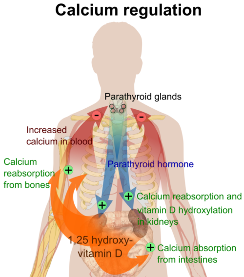
The major difference between anatomic compartments and physiologic function compartments is that anatomic compartments have physical borders and physiologic compartments are often amorphous – that is they do not occupy a fixed space. Another way of stating this is that anatomic compartments occupy space much like a room in a house occupies space. In contrast, the main characteristic of physiologic function compartments is that they are in a state of constant motion through space and over time. For example, physiologic functional compartments include the immune cell compartment, the trace metal compartment, the calcium ion compartment, and the extracellular fluid compartment.
Efficiency is gained by partitioning the body’s functional activities into compartments. By doing this, identical biological molecules can have many different functions depending upon their location in the internal milieu. For example, the molecule vasointestinal peptide (VIP) works in the gut to cause dilation of blood vessels, stimulates contractility of the heart, increases gluconeogenesis in the liver, lowers arterial blood pressure, relaxes smooth muscle of the trachea, stomach and gall bladder and regulates release of a pituitary hormone. Much of physiology is learning about how various functional compartments maintain their integrity and communicate with each other.
Microanatomy and compartments
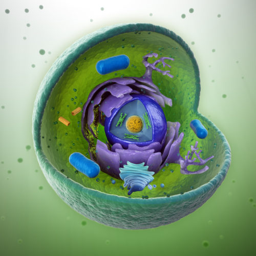
Most anatomical compartments are separated from each other by membranes composed of fat and protein. For example, fluids of the body are divided into two compartments, intracellular fluids found within cells and extracellular fluids outside cells. Cells themselves have many internal microanatomy compartments, the nucleus where the genetic code DNA is stored, mitochondria where energy from food is captured and a host of others.
Theories of physiology strive to accurately describe possible cross talk between the body’s micro compartments. This is where the facts keep changing. Scientists’ best story today about the details of these interactions will be tweaked when new data emerges from experiments in the near and far future.
To help you get a feel for the various levels at which compartments are studied, the following is a partial and representative list. A cell is a compartment. The nucleus of a cell is another compartment. There is the blood cell generating compartment in the marrow of bone. Also, there are the mineral compartments of the body. For example, the iron compartment of the human body includes enzymes, muscle, liver, red blood cells, and intestine.
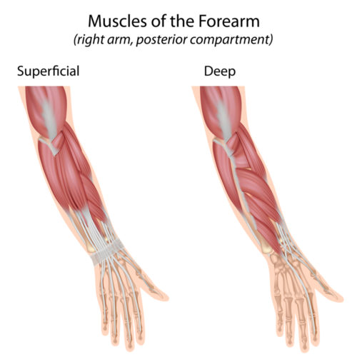
Muscles of the arms and legs are separated into isolated compartments with their own nerves and blood supplies by dense sheets of collagen material named fascia. The respiratory system has multiple anatomic and functional compartments. As you study physiology, remember that what you want to get your mind around are the limits of each compartment, and possible ways that communication can take place between body parts within those limits. If the boundary is a cell membrane, then ask what can pass through that membrane, why do the materials passing through travel in a particular direction, is cooperation between two or more molecules necessary for the passage of any of them to occur?
If you can begin to think in this way rather than trying to randomly memorize abstract theories, you will gain an understanding comparable to scientists working today in human physiology. There is a famous quote worth remembering by Carl Sagan, Scientist, 1934-1996. “Science is a way of thinking much more than it is a body of knowledge.”
Further Reading
Do you have questions?
Please put your questions in the comment box or send them to me by email at DrReece@MedicalScienceNavigator.com. I read and reply to all comments and email.
If you find this article helpful share it with your fellow students or send it to your favorite social media site by clicking on your favorite social media button.
Margaret Thompson Reece PhD, physiologist, former Senior Scientist and Laboratory Director at academic medical centers in California, New York and Massachusetts is now Manager at Reece Biomedical Consulting LLC.
She taught physiology for over 30 years to undergraduate and graduate students, at two- and four-year colleges, in the classroom and in the research laboratory. Her books “Physiology: Custom-Designed Chemistry”, “Inside the Closed World of the Brain”, and her online course “30-Day Challenge: Craft Your Plan for Learning Physiology”, and “Busy Student’s Anatomy & Physiology Study Journal” are created for those planning a career in healthcare. More about her books is available at https://www.amazon.com/author/margaretreece. You may contact Dr. Reece at DrReece@MedicalScienceNavigator.com, or on LinkedIn
Dr. Reece offers a free 30 minute “how-to-get-started” phone conference to students struggling with human anatomy and physiology. Schedule an appointment by email at DrReece@MedicalScienceNavigator.com.


I am a writer. I post essays on my site. I have written about cancer, and I am putting together another essay and am talking about cell physiology and the need to understand it, and I would like to use your graphic drawing of a cell. May I please use this drawing, it is well done.
Thank you. TJ
Hello Timothy. I do not own the picture of the cell. I licensed it for use from an online stock photo company at http://www.shutterstock.com. They have a large number of really good anatomy photos and vectors that they license royalty free for a small fee.
describe how molecules move within and between body compartments
For molecules to move between compartments there must be open channels in the compartment’s membrane through which molecules can move. The design of such channels is specific for each type of molecule. For example, a potassium ion cannot pass through a membrane sodium ion channel. Much of physiology is concerned with regulation of membrane channels. The driving force on each molecule is determined by the concentration gradient of the molecule on each side of the membrane and any electrical charges in the vicinity.
How molecules move within and between body compartment