Brain research is experiencing a data explosion
Many popular beliefs about human brain function were considered fact just 10 years ago. But now because of the brain research data explosion of recent years, scientists’ view of how the brain functions is changing. There is a long list of myths about the brain circulating widely on social media and elsewhere.
As a physiologist with an interest in brain function, I think there are 5 major brain myths that are more than ready for retirement. Here they are.
- Left brain vs.Right brain
- Only need 10% of brain cells
- Adult brain neuron connections fixed
- Accuracy of eyewitness testimony
- Human brain computer
1. Left brain vs. right brain
The idea that the left hemisphere of the brain is the source of creative and artistic activities and the right brain is reserved for logic and mathematical calculations was extrapolated from early discoveries in the late 1800s. Lichtheim (1885), Wernicke (1874) and Broca (1861) and others described separate areas of brain cortex necessary for language and speech in the left hemisphere.
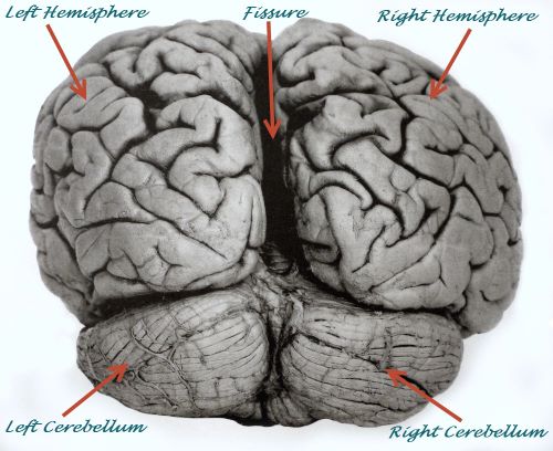
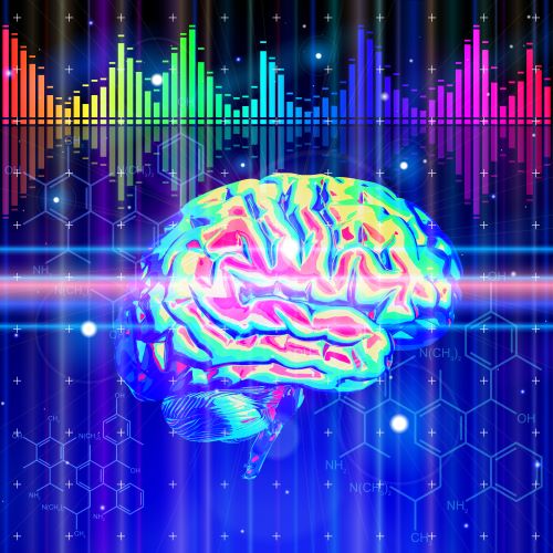
Human brain showing left and right hemispheres, cerebellum and central fissure, Pete Spiro/Shutterstock.com
The knowledge that language and speech required distinct regions of the left brain hemisphere emerged from patients in the clinic who suffered damage to those specific regions. While these patients contributed greatly to understanding brain cortical areas devoted to use of language, it is no longer believed that all aspects of speech and creative activity using words is managed by the language and speech centers in the left hemisphere.
Modern brain imaging studies discourage theories of complete dominance by one hemisphere for language and other brain functions. For example, recent modeling of neural pathways involved in hearing, language, speech production and speech recognition demonstrate that coordination of multiple bits of information are necessary. The multiple bits of information are spread over diverse brain regions in both hemispheres.
Hearing and speech recognition require many separate tasks with varying time course. Ability to accomplish the tasks is spread over a complex set of neural networks in both hemispheres. Cortical neurons and the neurons of deep brain nuclei all play their role. Sorting out all this activity is presently an active area of research.
The two hemispheres of the human brain are connected to each other by a bridge of neuron axons called the corpus callosum. Neuron cell bodies associated with these axons reside in both hemispheres. The axonal connections in both directions permit the right and left hemisphere to co-ordinate multiple complex logic and creative activities simultaneously.
2. Only need 10% of brain cells
The myth that only 10% of brain cells are needed comes from a fascination with the superstar cells of the brain, the neurons. In truth, neurons only represent about 10% of the brain’s cellular population. Thus, the idea arose that only 10% of the brain is required for mental function.
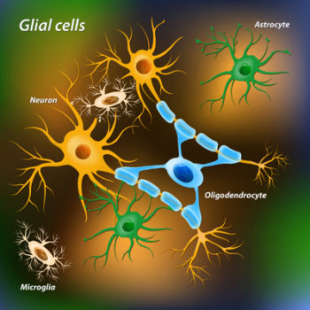
The names of the remaining 90% of brain cells are astrocyte, oligodendrocyte, microglia and ependymal cell. These non-neuron cells of the brain are generally known as glia. The name glia translates to mean ‘glue’. A major misnomer.
Early neuroanatomists, fascinated with the elaborate shape of the neurons, believed the more common appearing cells to be scaffolding for the neurons. In defense of these scientists of the early 20th century, their tissues staining methods detected only a small portion of the brain cell population, including only 1%-3% of the neurons.
Now it is known that astrocytes participate in neuron signaling at synapses. They form networks that separate neuron ensembles dedicated to specific tasks. Astrocytes also carry out parts of neurons’ nutrient metabolism to enhance energy availability for neurons. This allows neurons to rely entirely on glucose for energy.
Microglia monitors neuron synapse activity and repairs damaged neurons. If a neuron cannot be repaired microglia, with the help of astrocytes, removes the fatally damaged neuron. Microglia and astrocytes work together to coordinate efforts to fight infection and repair damaged areas of the brain.
Oligodendrocytes influence signaling along neuron axons. Oligodendrocytes wrap themselves around the axons of highly active neurons creating the white color of the brain’s nerve tracts. Electrical signals travel faster along axons coated intermittently with oligodendrocytes.
Transport systems for molecules moving to and from a neuron’s cell body along an axon depend on growth factors secreted by the oligodendrocytes. Pattern variation in the placement of oligodendrocytes along an axon affects neuron circuity, because axon synapses can only occur on bare patches of axon.
Ependymal cells line the hollow cavities in the brain. They produce the cerebral spinal fluid that brings nutrients into the brain and takes waste products out of the brain and into the general circulation for removal.
Without the glia and microglia, a brain could not function. There are about 86 billion neurons in the human brain. Astrocytes outnumber neurons by over 5 times and there is approximately 1 microglia per neuron. Oligodendrocytes and ependymal cells account for the remaining 90% of brain cells.
3. Adult brain neuron connections fixed
The first indication that “adult neuron connections are fixed” was a false idea came from research of songbird brains in the late 1970s. The way brain neurons connected in songbirds altered during the mating season to produce species specific songs associated with bird reproduction.
Dogma that the brain is hard-wired in adult humans survived until 1998 when it was shown that a region of cerebral cortex called the hippocampus could generate new neurons, supposedly from an adult neural stem cell population. This observation came from a pathology tissue section from the brain of a cancer patient who had been treated with a drug that only incorporates itself into DNA of newly made cells. Finding neurogenesis in the brain of an adult human opened a new era in brain research.
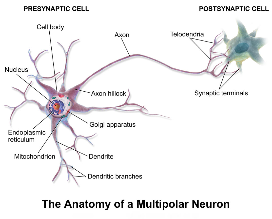
Scientists began to look more closely at the places on neurons where other neurons connect, structures named synapses. Neuron synapses occur on dendrites, the neuron cell body, axons and nerve endings. Some neurons even split their axons and send divisions, called collaterals, back to synaptic structures on their own cell body and/or dendrites.
Research of the last 10-15 years discovered that the synapses frequently active increase in size and multiply. Synapses that are used less often shrink and are pruned away by microglia. The coming and going of synaptic structures has a profound effect on the circuitry of adult brain neurons. It modifies the wiring pattern of the brain continually in response to new experiences. The name for this re-wiring process is neuroplasticity.
4. Accuracy of eyewitness testimony
Reports of eyewitnesses to an event carry a lot of weight when trying to sort out what happened at a later time. But the accuracy of eyewitness testimony in court has come into question. While eyewitnesses intuitively appear to be a valid approach to explaining past events, the way human memory is formed and recalled subverts the accuracy of the process. This form of memory is called episodic memory, and it is much like trying to remember who gave me which kernel of advice at my graduation party.
The form of memory that recalls specific events, situations and personal experience was named episodic memory by scientists who study human memory. Episodic memories are first coded by a region of the cerebral cortex called the hippocampus, the only region of the brain thought to be able to produce new neurons through decades of life.
Hippocampus memory codes are divided into pieces and the fragments move to separate locations throughout the brain for storage. Visual representation, auditory representation and emotional representation may all map to different brain regions. To recall a memory the brain must reassemble its parts.
The act of retrieving an episodic memory reshapes pieces of the older memory into a new episodic memory. Old memories become updated with each recall to include new experience. In this way the brain integrates what is already understood to be true with everything new it learns. This updating of memory to include additional information at each recall makes eyewitness accounts of past events unreliable.
5. Human brain computer
Digital computers are getting better at simulating tasks accomplished by the human brain, but they still have a long way to go to match the analytic power of the human brain. Brains solve multiple problems and engage in complex environmental analysis all at the same time. They do this with little error even though it is a watery structure tens of millions of times smaller and slower than state-of-the-art digital computers.
Computer signaling follows a fixed digital on/off sequential program. Signaling in the brain is no longer thought to be strictly digital or sequential. Rather the brain uses ensemble coding by groups of neurons working in parallel to eliminate noise in its electrochemical circuity. Signals are sent through relay hubs much like the flight paths of airlines are setup through hub cities. There is more than one way to get from point A to point B.
Interestingly, ensemble groups are not static in their composition. The number of participating neurons and the neurotransmitters chosen for use vary over time. It was once thought that each neuron’s transmitter molecule was fixed. Now it is known that a single neuron can change its neurotransmitter depending upon incoming environmental signals.
There is also activity-dependent recruitment of neurons from reserve neuron pools to ensemble circuits. The reserve-pool neurons share input and output with the core ensemble. Reserve-pool neurons may or may not use a different neurotransmitter than the core group to affect common targets. Reserve neuron pools working with core ensembles are observed across vertebrate brains from frogs to primates.
Even with ensemble coding, information moves quicker through the brain than its chemistry suggests it should. One theory is that the balance between stimulatory and inhibitory circuit activity, which causes electrical rhythms in the brain, may align information coming in from multiple sensory systems with neuron ensemble activity to produce one overall message to widespread cortical neurons.
If you find interesting the retirement of brain myths with the increase in brain research, you can discover more about how the human brain really functions in my book “Inside the Closed World of the Brain, How brain cells connect, share and disengage–and why this holds the key to Alzheimer’s disease.” Click here for a look inside.
More about the brain:
2017 Virtual Tour Inside the Closed World of the Brain
Hearing: Sensing the World Through Sound Waves
Birth of New Neurons Continues into the 5th Decade
Do you have questions?
Send me an email with your questions at DrReece@MedicalScienceNavigator.com or put them in the comment box. I always read my mail and answer it. Please share this article with your fellow students taking anatomy and physiology by clicking your favorite social media button.
 Margaret Thompson Reece PhD, physiologist, former Senior Scientist and Laboratory Director at academic medical centers in California, New York and Massachusetts is now Manager at Reece Biomedical Consulting LLC.
She taught physiology for over 30 years to undergraduate and graduate students, at two- and four-year colleges, in the classroom and in the research laboratory. Her books “Physiology: Custom-Designed Chemistry”, “Inside the Closed World of the Brain”, and her online course “30-Day Challenge: Craft Your Plan for Learning Physiology”, and “Busy Student’s Anatomy & Physiology Study Journal” are created for those planning a career in healthcare. More about her books is available at https://www.amazon.com/author/margaretreece. You may contact Dr. Reece at DrReece@MedicalScienceNavigator.com, or on LinkedIn.
Dr. Reece offers a free 30 minute “how-to-get-started” phone conference to students struggling with human anatomy and physiology. Schedule an appointment by email at DrReece@MedicalScienceNavigator.com.

