Dendritic spines & memory
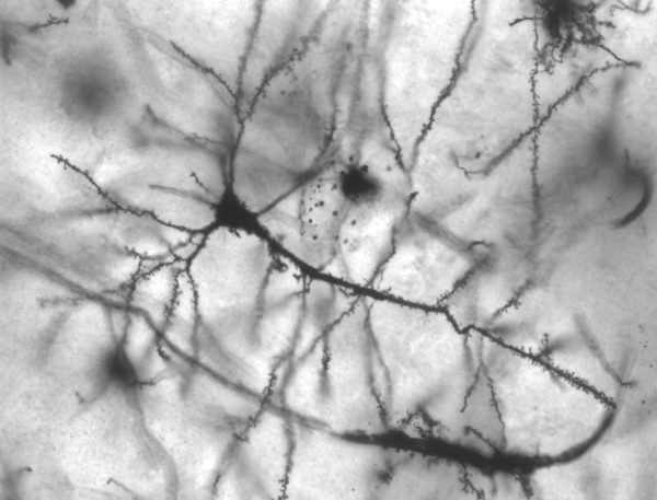
Dendritic spines are the anatomical form of memory. Anatomy and physiology lectures present a broad array of audio and visual cues that are aimed at helping your brain remember specific aspects of these sciences. The actual mechanism underlying your memory of these stimuli is best illustrated by the dendritic spine’s plasticity on brain neuron dendrites. The flexible anatomy of these very tiny brain compartments allows you to create memories that can be recalled and manipulated to answer questions on your exams.
There are two major types of memories. They are declarative memory and procedural memory. Each uses different brain pathways. Declarative or explicit memory includes memories of facts and knowledge. Procedural memory is an unconscious memory of how to do something like ride a bike, drive a car, type on a keyboard, text on a cell phone.
Hippocampus and memory
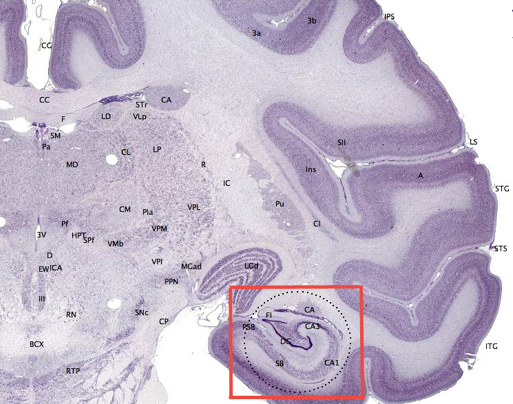
The hippocampus is the part of the brain where declarative memory begins. In this map of a monkey brain, the hippocampus is displayed within the red box. Any guesses where this brain structure got its name? Can you imagine the shape of a seahorse there? The organization of the area is anatomically similar in humans.
Declarative memory is the type of memory you are trying to enhance when you study anatomy and physiology. Because the physiology of memory construction is conserved across species most mechanistic studies of memory have been accomplished using rats and monkeys.
When sensory organ output pathways send excitatory electrical signals to hippocampus neurons, the incoming sensory neuron terminals search for spines on dendrites where they can build specialized connections called synapses. Because memory formation is an ongoing dynamic brain function, dendritic spines density is studied most thoroughly in the hippocampus.
Dendritic spine function
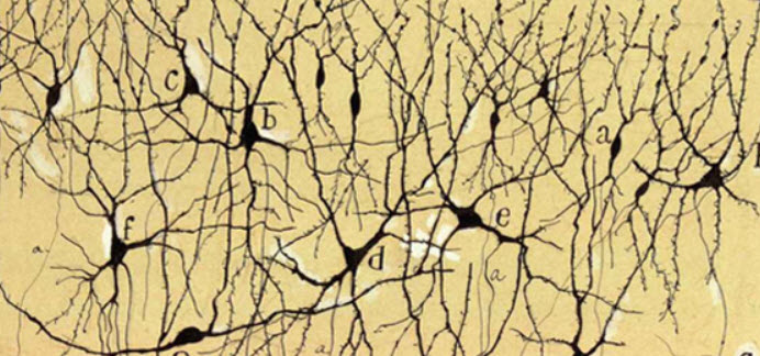
Dendritic spine function was a mystery until recently even though dendritic spines were present in drawings of brain neurons created over 100 years ago. This image is a drawing of hippocampus neurons stained with silver by Ramön y Cajal, a neuroscientist who won the Nobel Prize in 1906.
Brain dendritic spines with their neurotransmitter receptors are in a constant state of turnover. New spines form on brain dendrites, mature, and are pruned away by microglia in the adult brain and in the developing brain of children. The number, shape, and size of dendritic spines varies in direct correlation with the magnitude of sensory input. Average spine turnover rates have been reported to be as high as 6-15% per day.
Yet, it has only been in the past 10 years that technological developments permitted detailed study of the relationship between structure and dendritic spine function. Developments in fluorescent microscopy of the brains of living animals have permitted demonstration of the autonomy of dendritic spines as functional compartments within neurons.
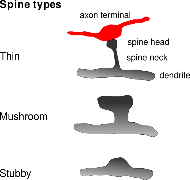
Synapses on dendritic spines in the hippocampus that receive high frequency auditory or visual stimuli become stronger. Increased spine strength correlates with larger spine size. The enlargement of the spine is due to an increase in new protein synthesis by the neuron cell body to better manage excitatory incoming traffic.
Spine enlargement is tightly linked to an increase in intracellular calcium concentration within the spine. Dendritic spines where calcium remains low due to lack of neuronal signaling are pruned from the dendrite by microglia.
Increased calcium in the dendritic spine promotes synthesis of molecules that travel to the neuron’s nucleus and turn on synthesis of new proteins for the synapse. Entire dendrites with a large percentage of inactive spines are also pruned from neurons.
Long term memory
The hippocampus is responsible for establishing long term memory. Consolidating a locally encoded dendritic spine memory for long-term use is a relatively slow process. It involves hippocampus driven transfer of the anatomical memory configuration to various locations in the brain for storage. Visual representation, auditory representation and the emotional components of an event may all map to different brain areas.
The act of retrieving an episodic memory reshapes pieces of the older memory into an updated memory that includes new experience with each recall. Human noninvasive brain imaging studies show that recall of a memory follows pathways that are the reverse of those used for memory formation and memory consolidation. If the pathway of memory formation is complex, the process of recall will be complex as well.
Thus, when learning something new, it is important to keep the learning process as organized as possible. Trying to learn many facts in a random fashion makes recall more difficult. The brain likes established patterns where it can tuck in new facts and experiences.
Dendritic spine function is one of the most active area of brain research. That dendritic spine morphology correlates with memory formation is a first clue about how long term memory and awareness happens.
Further reading:
Memory, Patterns, and Themes in Anatomy & Physiology
Do you have questions?
Please put your questions in the comment box or send them to me by email at DrReece@MedicalScienceNavigator.com. I read and reply to all comments and email.
If you find this article helpful share it with your fellow students or send it to your favorite social media site by clicking on one of the buttons below.
Margaret Thompson Reece PhD, physiologist, former Senior Scientist and Laboratory Director at academic medical centers in California, New York and Massachusetts is now Manager at Reece Biomedical Consulting LLC.
She taught physiology for over 30 years to undergraduate and graduate students, at two- and four-year colleges, in the classroom and in the research laboratory. Her books “Physiology: Custom-Designed Chemistry”, “Inside the Closed World of the Brain”, and her online course “30-Day Challenge: Craft Your Plan for Learning Physiology”, and “Busy Student’s Anatomy & Physiology Study Journal” are created for those planning a career in healthcare. More about her books is available at https://www.amazon.com/author/margaretreece. You may contact Dr. Reece at DrReece@MedicalScienceNavigator.com, or on LinkedIn.
Dr. Reece offers a free 30 minute “how-to-get-started” phone conference to students struggling with human anatomy and physiology. Schedule an appointment by email at DrReece@MedicalScienceNavigator.com.

