Oligodendrocytes function

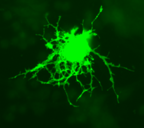
Oligodendrocyte in culture (mouse) transfected with Green Fluorescent Protein 630X, Jurjen Broeke/Wikimedia Commons
Neuron myelination
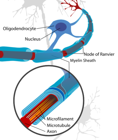
Oligodendrocytes are the glial cells in the brain responsible for neuron myelination. Like other glial cells, oligodendrocytes possess a cell body (soma) from which many processes extend. Extension and retraction of oligodendrocyte processes are crucial for their migration through the brain and establishing their contact with neurons.
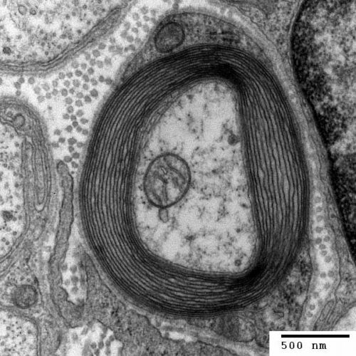
The membrane of the oligodendrocyte processes has a high content of lipid bound protein called lipophilin. Upon contact with neurons, oligodendrocyte exploratory processes convert to flat sheets that spread and wind around the axons to create a multilayered or laminar cover. The laminar structure then compacts extruding almost all its cytoplasm and is referred to as myelin. Oligodendrocytes select larger diameter axons for myelination. A single oligodendrocyte may envelop as many as 6o axons simultaneously.
Neuron conduction velocity
Neuron conduction velocity depends upon axon myelination. Myelination of brain neurons by oligodendrocytes enables rapid propagation of electrical signals, action potentials, along neuronal axons. Myelination increases conduction velocity of axons by at least 50 times.
Besides increasing neuron conduction velocity, myelin also nourishes and protects neuron axons against inflammatory and oxidative injury by placing multiple layers of lipid between axons and highly reactive oxygen-containing molecules. The human brain, and that of other mammals, is characterized by a high rate of production of reactive molecules. Destruction of the myelin sheath around neuron axons rapidly leads to neuron death.

Cross section of a neuron axon with its myelin sheath, Generated and deposited into the public domain by the Electron Microscopy Facility at Trinity College/Wikimedia Commons
There is evidence that myelination of neurons is partly driven by the level of electrical activity in axons themselves. For example, changes in neuronal activity can increase myelination of neurons in the motor cortex when new learning involves intricate motor skill patterns requiring speed and precision, such as playing musical instruments.
A suggested mechanism for this process is hypothesized to involve axonal release of ATP that stimulates adjacent astrocytes to release the pro-myelination cytokine LIF, which in turn signals to oligodendrocytes to increase the level of myelination.
Brain myelination time course
The time course of brain myelination extends through most of a lifetime. While myelination of neurons in the human brain starts in late fetal life and peaks in infants, certain associative regions continue to increase brain myelination into the 5th and 6th decade of life.
White matter, neuron axons covered by myelin, of the adult brain is densely populated with both mature oligodendrocytes and oligodendrocyte precursor cells. Regeneration of brain myelination after injury depends upon activation of the widespread pool of oligodendrocyte precursor cells.
Many cellular factors and extracellular signaling molecules have been implicated in maturation of oligodendrocyte precursor cells. An excellent review of this process by Ben Emery is available at Science, VOL 330, 2010, pages 779-772.
Destruction of myelin sheath
Several disease processes in humans include destruction of the myelin sheath of neurons in the brain and spinal cord. Among these are Multiple Sclerosis (MS) and Alzheimer’s disease.
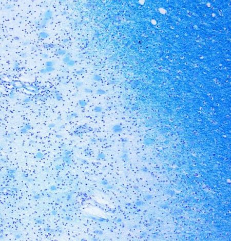
MS is an inflammatory disease in which myelin around axons of the brain and spinal cord are damaged and scarred. In MS the myelin destruction becomes progressively widespread throughout the central nervous system. The precise cause of MS is still unknown, but it is thought that the oligodendrocytes die.
Klüver-Barerra Stain colors myelin in brain tissue dark blue. The light blue area of this photomicrograph is a demyelinated MS lesion in the brain with adjacent normal tissue to the right.

Photomicrograph of a demyelinating MS-Lesion in the brain, Klüver-Barerra-Stain at 10X,, Marvin 101/Wikimedia Commons
In contrast, demyelination of axons in Alzheimer’s, a form of dementia associated with brain inflammation, is a more discrete process. In Alzheimer’s there is a focal loss of oligodendrocytes associated with amyloid beta plaque. Plaque free regions display little to no demyelination or oligodendrocyte loss.
Do you have questions?
Please put your questions in the comment box or send them to me by email at DrReece@MedicalScienceNavigator.com. I read and reply to all comments and email.
If you find this article helpful share it with your fellow students or send it to your favorite social media site by clicking on one of the buttons below.
Further reading
Neurons: Where Does Their Electricity Come From
Neurons and Brain’s Other 90% of Cells
Margaret Thompson Reece PhD, physiologist, former Senior Scientist and Laboratory Director at academic medical centers in California, New York and Massachusetts is now Manager at Reece Biomedical Consulting LLC.
She taught physiology for over 30 years to undergraduate and graduate students, at two- and four-year colleges, in the classroom and in the research laboratory. Her books “Physiology: Custom-Designed Chemistry”, “Inside the Closed World of the Brain”, and her online course “30-Day Challenge: Craft Your Plan for Learning Physiology”, and “Busy Student’s Anatomy & Physiology Study Journal” are created for those planning a career in healthcare. More about her books is available at https://www.amazon.com/author/margaretreece. You may contact Dr. Reece at DrReece@MedicalScienceNavigator.com, or on LinkedIn.
Dr. Reece offers a free 30 minute “how-to-get-started” phone conference to students struggling with human anatomy and physiology. Schedule an appointment by email at DrReece@MedicalScienceNavigator.com.


